ELECTRON MICROSCOPY × PROTEIN DESIGN
See what you made.
Jellyfold provides focused consulting at the intersection of protein biochemistry, electron microscopy, and computational protein design.

THE JELLYFOLD POINT OF VIEW
Electron microscopy is part of the protein design pipeline.
Jellyfold believes EM belongs within the protein design process, not downstream of it or separate from it. Bringing structural evidence into the pipeline helps protein designers make better decisions, iterate more effectively, and move forward faster.
This does not always require high-resolution cryo-EM. Negative-stain EM is an accessible and often overlooked tool that produces structural data quickly and makes it easier to assess designs throughout the design cycle.
THE CONFIDENCE CASE
When it works, prove it.
Give a promising computational design the structural validation it deserves.
WHEN THE DESIGN DELIVERS
A confident final structure drives the point home.
When the biology looks promising, EM data can show whether your designed structure is on target—and explain why. Use that evidence to strengthen a publication, support a patent, build an approval package, or guide the next design decision.
WHERE I CAN HELP
Electron microscopy consulting for the protein design cycle.
WHEN DESIGN MEETS REALITY
The design looked right. Until it didn’t.
We've all been there. Your design agrees with leading folding predictors. Then the wet-lab results are strange: activity is missing, binding is weak, or behavior doesn’t match the hypothesis.
Is the functional hypothesis wrong, or did the protein fail to fold or assemble as intended?
THE STRUCTURAL FEEDBACK LOOP
Catch problems early.
Jellyfold helps you avoid common experimental-validation pitfalls and use structural evidence to assess whether a design folds, assembles, and behaves as intended. Apply that evidence early and often to guide iteration before weak candidates consume more time and resources.
See what you made.
WHY JELLYFOLD
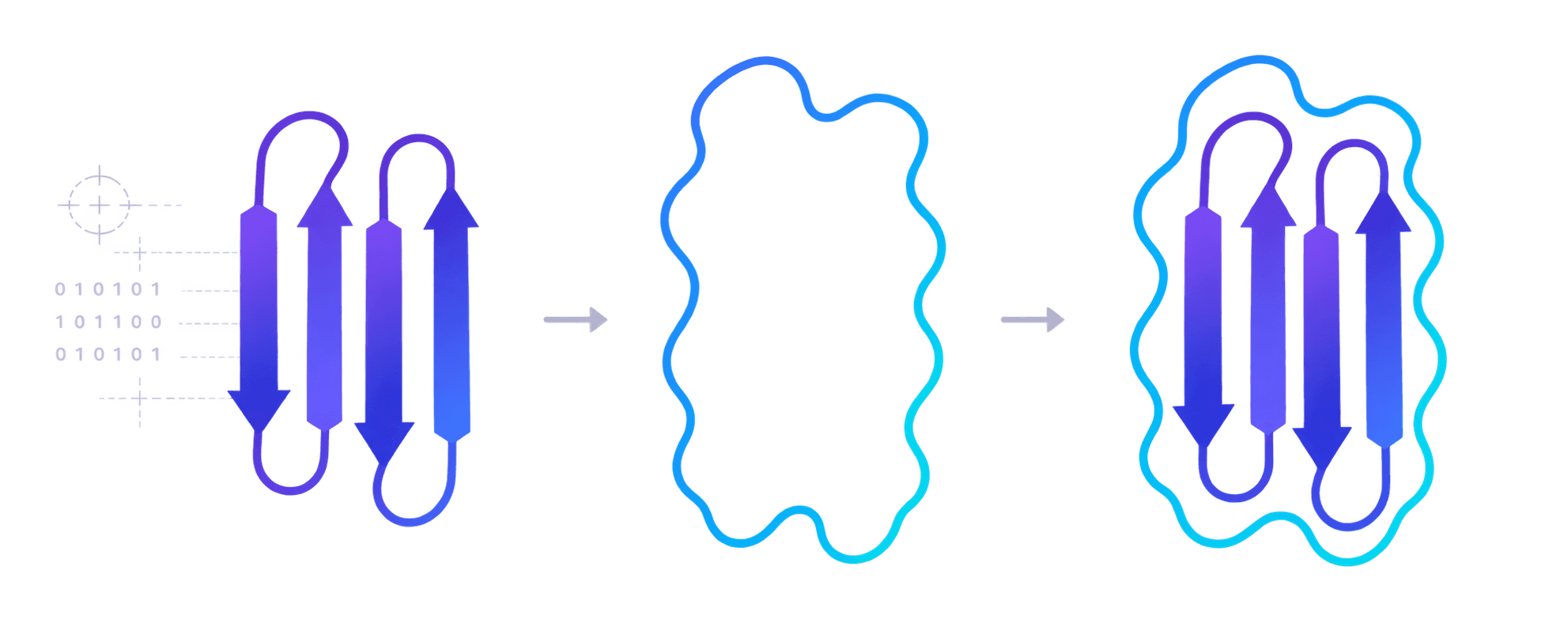
The fold within the density.
WHAT THE MAP CAN SUPPORT
01
Domain level: is the overall design architecture and assembly state on target?
02
Backbone level: does the experimental structure match the designed protein fold?
03
Side chains level: are key rotamers positioned to support the expected interactions?
04
Atomic level: does the map resolve atomic-level interactions that explain function?
ABOUT ANDREW
Structural biology expertise, built for the protein design loop.
Andrew J. Borst, Ph.D., is a structural biologist and electron microscopy leader with deep experience in protein biochemistry, biophysics, negative-stain EM, cryo-EM, and computational protein design. At the University of Washington’s Institute for Protein Design, he leads the Electron Microscopy R&D Core, building high-throughput pipelines for thousands of protein design targets and helping protein design scientists learn and use EM to answer structural questions throughout the design cycle. His work spans de novo binder design, designed protein assemblies, antibody–antigen complexes, computationally stabilized antigens, and nanoparticle vaccine platforms, including structural characterization supporting RFdiffusion and RFantibody, with a focus on translating experimental structure into clear guidance for computational protein design.

15
Years of EM experience
40+
Cryo-EM structures determined
1,000s
Designed protein samples characterized by negative-stain EM
120+
Protein design scientists mentored and trained in EM
LET’S TALK
Have a structural problem worth untangling?
Tell me what you’re working on. We’ll see whether Jellyfold is a fit for your research, team, or business.